
Modern healthcare increasingly relies on technology to help doctors identify medical conditions more accurately and make informed treatment decisions. This progress is especially important in vascular care, where understanding how blood moves through the veins can help explain symptoms that may otherwise go unnoticed. For people experiencing leg swelling, discomfort, or visible changes in their veins, access to proper evaluation and advanced vein treatment in Taylor can be an important part of managing their condition.
Vein disorders can develop gradually, and their symptoms may be mistaken for ordinary tiredness or the effects of spending too much time sitting or standing. Modern diagnostic methods give healthcare professionals better ways to examine circulation, identify possible problems, and determine which treatment approaches may be appropriate.
Understanding Common Vein Disorders
Veins are blood vessels responsible for carrying blood back toward the heart. In the legs, this process works against gravity, so small one-way valves help keep blood moving in the right direction.
When these valves become weak or damaged, blood may flow backward and collect in the veins. This condition is known as venous reflux. Over time, increased pressure can cause veins to become enlarged and twisted, resulting in varicose veins.
Other venous conditions include chronic venous insufficiency, which occurs when the veins have difficulty returning blood efficiently to the heart. People with this condition may experience swelling, aching, skin discoloration, or changes around the ankles and lower legs.
Some symptoms are visible, while others are less obvious. This is why examining the underlying blood flow is often an important part of the diagnostic process.
The Role of Technology in Modern Vein Diagnosis
Technology has changed the way healthcare professionals investigate vein-related problems. Instead of relying only on visible symptoms and physical examinations, doctors can use imaging tools to study the structure and function of blood vessels.
These methods provide additional information about circulation, helping clinicians understand whether symptoms may be connected to venous reflux, obstruction, or another condition.
Duplex Ultrasound Imaging
Duplex ultrasound is one of the most widely used diagnostic tools for evaluating vein disorders. It combines traditional ultrasound imaging with Doppler technology to examine both the structure of blood vessels and the movement of blood through them.
During the examination, a technician or qualified healthcare professional moves a handheld device over the skin. The device uses sound waves to produce images of the veins and assess blood flow.
This test can help identify enlarged veins, abnormal blood movement, and problems with the valves that normally prevent blood from flowing backward.
One major advantage of duplex ultrasound is that it does not require surgery or exposure to ionizing radiation. It is also generally comfortable and can provide useful information during an outpatient appointment.
The results can help a specialist determine the location and extent of a vein problem before discussing possible treatment options.
Doppler Ultrasound Technology
Doppler ultrasound focuses on measuring the movement of blood through vessels. It uses changes in sound-wave frequency to provide information about the direction and speed of blood flow.
This is particularly useful when a doctor needs to investigate whether blood is moving normally through the veins or flowing backward because of faulty valves.
The information gathered through Doppler imaging can support the assessment of venous insufficiency and other circulation-related concerns.
Although Doppler technology is often included in duplex ultrasound examinations, the two terms describe different aspects of the imaging process. Duplex ultrasound combines anatomical imaging with Doppler-based blood-flow assessment.
High-Resolution Imaging
Improvements in ultrasound equipment have made it possible to produce clearer images of blood vessels and surrounding structures. Better image quality can help healthcare professionals examine smaller details and identify abnormalities that may be difficult to assess through a physical examination alone.
High-resolution imaging also supports more detailed mapping of the affected veins. This information can be useful when planning procedures that target specific sections of a vein.
However, the quality of a diagnosis depends on more than the equipment itself. The experience of the professional performing and interpreting the examination remains important.
How Accurate Diagnosis Supports Better Treatment Decisions
Identifying a vein condition is only the first step. Healthcare professionals also need to understand its location, severity, and possible causes before recommending an appropriate care plan.
Diagnostic imaging helps provide this information.
For example, two people may both have visible varicose veins but experience different symptoms. One person may mainly be concerned about the appearance of the veins, while another may experience persistent heaviness, swelling, or discomfort.
An ultrasound examination can help determine whether abnormal blood flow is contributing to these symptoms. It can also help identify which veins are affected and whether additional evaluation is necessary.
This information allows doctors to consider treatment options based on the individual's condition rather than relying on appearance alone.
Modern Treatment Options for Vein Disorders
Advances in diagnosis have also supported the use of several minimally invasive treatments for selected vein conditions. The most suitable option depends on the patient's medical history, examination findings, symptoms, and ultrasound results.
Endovenous Laser Treatment
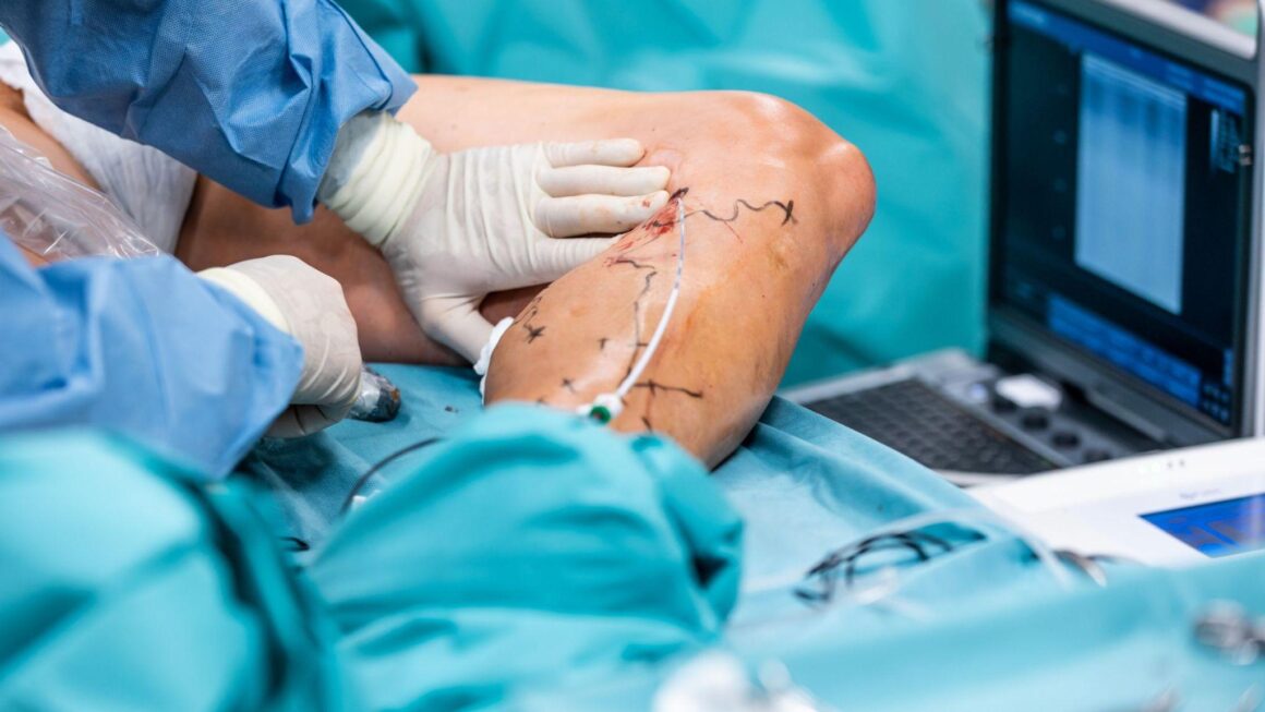
Endovenous laser treatment uses laser energy to close an affected vein from the inside. A thin catheter is guided into the targeted vein, and controlled heat is delivered through a laser fibre.
The treated vein gradually closes, allowing blood to flow through healthier nearby veins.
The procedure is generally performed using local anesthesia and is often considered for patients with certain types of venous reflux. A specialist determines whether it is suitable after reviewing the diagnostic findings.
Radiofrequency Ablation
Radiofrequency ablation is another minimally invasive method used to treat selected veins with faulty valves. It uses controlled radiofrequency energy to heat and close the affected vein.
Similar to laser treatment, the procedure is performed through a small access point rather than requiring a large surgical incision.
Both laser and radiofrequency procedures aim to redirect blood flow through healthier veins. However, they are not appropriate for every patient, and individual evaluation is necessary before choosing between them.
Sclerotherapy
Sclerotherapy is commonly used for certain smaller varicose veins and spider veins. During the procedure, a healthcare professional injects a solution into the targeted vein. This causes the vein to close over time.
The treated vessel gradually becomes less visible as the body absorbs it. Depending on the size and location of the veins, more than one treatment session may be needed.
The type of sclerotherapy used and the expected results depend on the individual case.
Ambulatory Phlebectomy
Ambulatory phlebectomy is a procedure used to remove selected surface varicose veins through very small skin openings. It may be recommended when enlarged veins are close to the surface and are suitable for removal.
The procedure is generally performed under local anesthesia. The specialist removes the targeted veins through small incisions, which typically do not require traditional stitches.
A doctor will determine whether this option is appropriate based on the vein's location and the patient's overall condition.
Why Personalized Evaluation Matters
Technology provides valuable information, but it does not replace professional judgment. A diagnosis should consider the patient's symptoms, medical history, physical examination, and imaging results together.
For example, leg swelling can be associated with several health conditions, not just vein disorders. A specialist may need to consider other possible causes before recommending treatment.
Personalized evaluation also helps prevent unnecessary procedures. Some people with visible veins may not require medical intervention, while others with less obvious changes may have symptoms linked to underlying venous reflux.
A clear discussion with the healthcare provider can help patients understand their diagnosis, available options, potential risks, and expected recovery process.
What Patients Can Expect During a Vein Evaluation
A vein assessment usually begins with a discussion about symptoms and medical history. Patients may be asked when the discomfort started, whether swelling becomes worse during the day, and whether they have a family history of vein conditions.
The healthcare professional may then examine the legs for visible veins, swelling, skin changes, or other signs that require further investigation.
If necessary, an ultrasound examination may be arranged to evaluate blood flow and valve function. The findings help the specialist explain whether a vein disorder is present and what options may be suitable.
Patients should feel comfortable asking questions about the diagnostic process, the purpose of recommended tests, and the differences between available treatments.
The Future of Technology in Vascular Care
Medical technology continues to develop, creating new opportunities to improve the way vascular conditions are identified and managed. Better imaging systems, improved procedural equipment, and more detailed diagnostic information may help healthcare professionals plan care with greater precision.
Digital medical records and improved imaging storage can also make it easier to review previous examinations and monitor changes over time.
However, technological progress should always support good clinical practice. Equipment alone cannot guarantee an accurate diagnosis or successful treatment. Proper training, careful interpretation, and communication between patients and healthcare professionals remain essential.
As these tools continue to improve, patients may benefit from more informed discussions and treatment plans that reflect their individual needs.
Conclusion
The diagnosis of vein disorders has become more detailed with the development of modern imaging technology. Duplex ultrasound, Doppler assessment, and improved imaging equipment allow healthcare professionals to examine blood flow, identify abnormal valve function, and understand the extent of venous disease.
These diagnostic tools also support more informed treatment decisions, including the use of minimally invasive procedures when appropriate. For patients experiencing persistent leg discomfort, swelling, or visible vein changes, seeking a professional evaluation can help identify the cause and clarify the next steps.
Ultimately, combining modern technology with experienced medical care gives patients a better foundation for understanding their vein health and making informed decisions about treatment.